
Fig. 4:
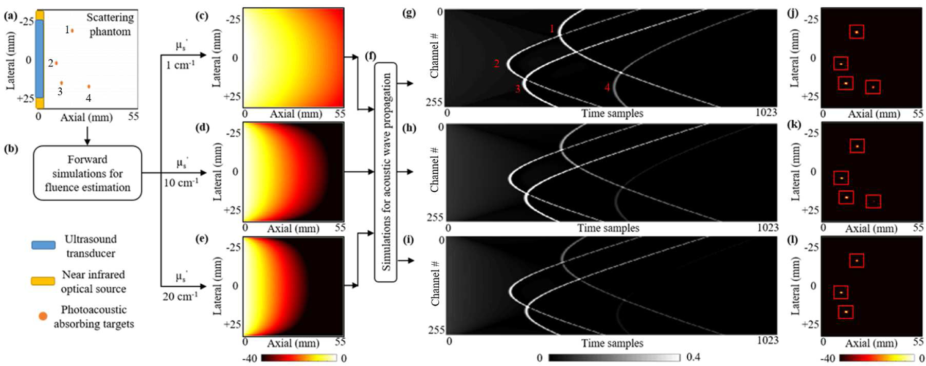
The schematic of our simulated dataset generation platform with different optical scattering levels and numbers of targets. (a) A schematic view of the 55×55 mm digital tissue phantom. A 51.2 mm wide 256-element ultrasound transducer array (blue stripe) and a 54 mm wide optical source (yellow stripe) are placed along the left edge of the phantom. Orange circles are 0.3 mm diameter vascular targets. (b) Simulated diffused light propagation via the NIRFAST toolbox solves optical fluence distributions for three different tissue mediums with reduced optical scattering coefficients of (c) 1 cm−1; (d) 10 cm−1; and (e) 20 cm−1. (f) The photoacoustic wave propagation resulting from the optical fluence-induced initial pressure is simulated via the K-Wave toolbox. (g, h, i) The time sampled photoacoustic signals detected by the 256-element transducer array for each scattering level and (j, k, l) their corresponding beamformed images.