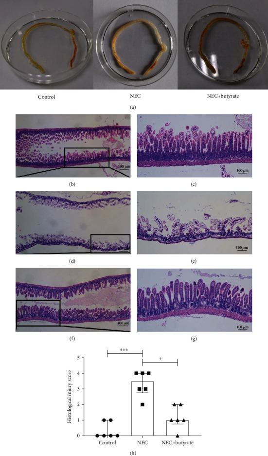
Figure 2.

(a) Visual morphological observation of intestinal tissue from newborn mice in the three groups. (b–g) Histopathological observation of the terminal ilea in the three groups; images of HE staining observed by microscopy; (b, c) control group; (d, e) NEC group; (f, g) NEC+butyrate group. Magnification: 40x and 100x; scale bar = 100 μm. (h) The intestinal histopathological injury scores in the three groups. Number of samples: control (n = 6), NEC (n = 6), and NEC+butyrate (n = 6). Statistical analysis: Kruskal-Wallis test. ∗P < 0.05; ∗∗∗P < 0.001.
