Figure 6.

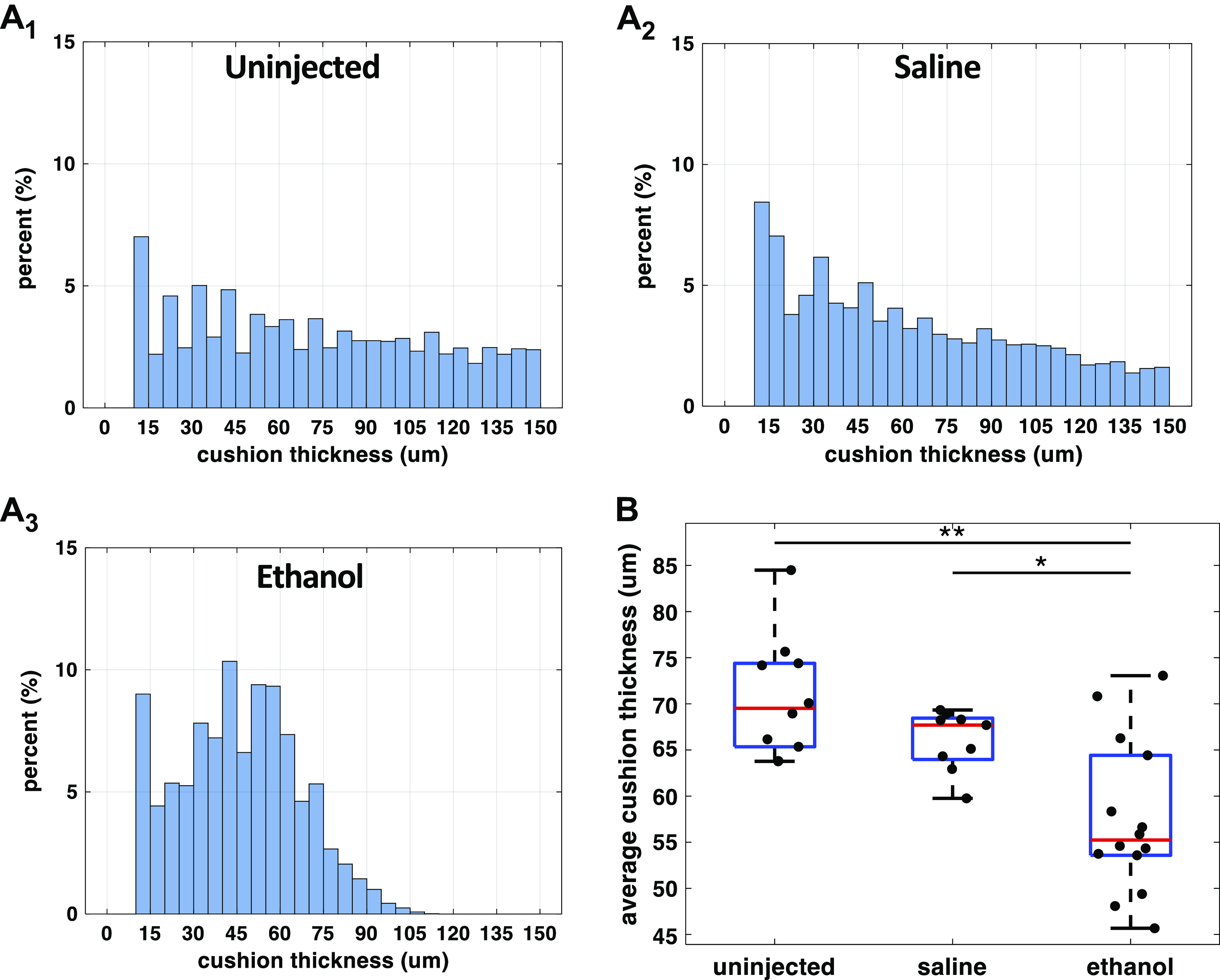
Ethanol exposure reduced endocardial AVJ cushion thickness at HH stage 20. A1–A3: cushion thickness histograms from three representative embryos: an untreated, a saline-treated, and an ethanol-treated embryo. At every point on the heart’s abluminal cushion surface, its distance to the luminal cushion surface was calculated as the cushion thickness at that location. From each heart, a range of cushion thickness values was generated. The y-axis of the histogram is the percentage of a specific thickness value in all measurements from one heart. B: quantification of endocardial cushion thickness among the three treatment groups: untreated (n = 10), saline-treated (n = 9), and ethanol-treated groups (n = 14). n, number of embryos. The ethanol group developed thinner cushions compared with controls (*P < 0.05, **P < 0.01). The comparison between the three groups was performed with one-way ANOVA and the Tukey–Kramer post hoc test. AVJ, atrioventricular junction; HH, Hamburger–Hamilton.
