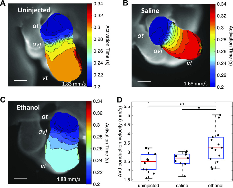
Figure 8.
Ethanol-treated embryos developed faster AVJ conduction at HH stage 20. A–C: activation time maps of embryonic hearts from three groups: untreated, saline-treated, and ethanol treated. Scale bar = 200 μm. D: quantification of conduction velocity at AVJ among the three treatment groups: untreated (n = 10), saline-treated (n = 9), and ethanol-treated groups (n = 14). n, number of embryos. The ethanol group developed faster conduction at AVJ compared with controls (*P < 0.05, **P < 0.01). The comparison between the three groups was performed with one-way ANOVA and the Tukey–Kramer post hoc test. At, atrium; AVJ, atrioventricular junction; HH, Hamburger–Hamilton; Vt, ventricle.

