Figure 2.
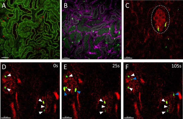
Intravital snapshots of the murine kidney cortex. A: neutrophils (red) are observed among spaces located between renal tubules (dull green, autofluorescence) in the kidney cortex. Some S1 and S2 renal tubules that can be differentiated as S1 proximal tubules have coarse green-red (yellow) autofluorescence vs. S2 with only fine green autofluorescence. During disease, these neutrophils would transmigrate from the capillaries into the kidney interstitium contributing to renal injury. Scale bar 50 μm. B: neutrophils (green) that are between renal tubules are located in the peritubular capillaries (magenta). Labeling with additional fluorescent antibodies against capillaries diminished the autofluorescence contrast between S1 and S2 renal tubules. Scale bar 50 μm. A and B were visualized with a Leica DMI6000 and Nikon Ti2 inverted spinning-disk confocal microscope, respectively, equipped with a ×20 Plan Apo objective lens (NA0.7 and NA0.8) on a C57Bl/6 mouse. Alexa Fluor (AF)594-conjugated and AF488-conjugated Anti-Ly6G (clone 1A8) were used to label neutrophils in A and B correspondingly. AF647 Isolectin B4 and Brilliant Violet 421 Anti-CD31 (clone 390) were used to label the endothelial cells in B, the acquired signals were combined into a single color (magenta) to highlight the endothelium. C: neutrophils (green) transiently present in the ball-like glomerulus (red, dotted white circle outline) labeled with AF594-conjugated 10 kDa dextran and visualized by a custom-made multiphoton microscope equipped with coherent tunable femtosecond Ti-Sapphire laser tuned to 780 nm (×20 water objective, NA0.95) on a Lysozyme M (LysM)-GFP mouse. Scale bar 50 μm. D–F: timelapse snapshots of neutrophils (green) in glomeruli (red) under 2 min, 2 glomeruli could be seen in this field of view. Some neutrophils persisted in the glomeruli during the imaging period (white arrows), whereas others were observed to rapidly flow through the glomeruli (blue arrows). During anti-GBM glomerulonephritis, these neutrophils significantly increase their dwell time within the glomeruli through the upregulation of adhesion factors and interaction with other immune cells and cause injury through release of reactive oxygen species, proteases, and NETs (Fig. 1B). Scale bar 50 μm. GBM, glomerular basement membrane; NETs, neutrophil extracellular traps.

