
Figure 1.
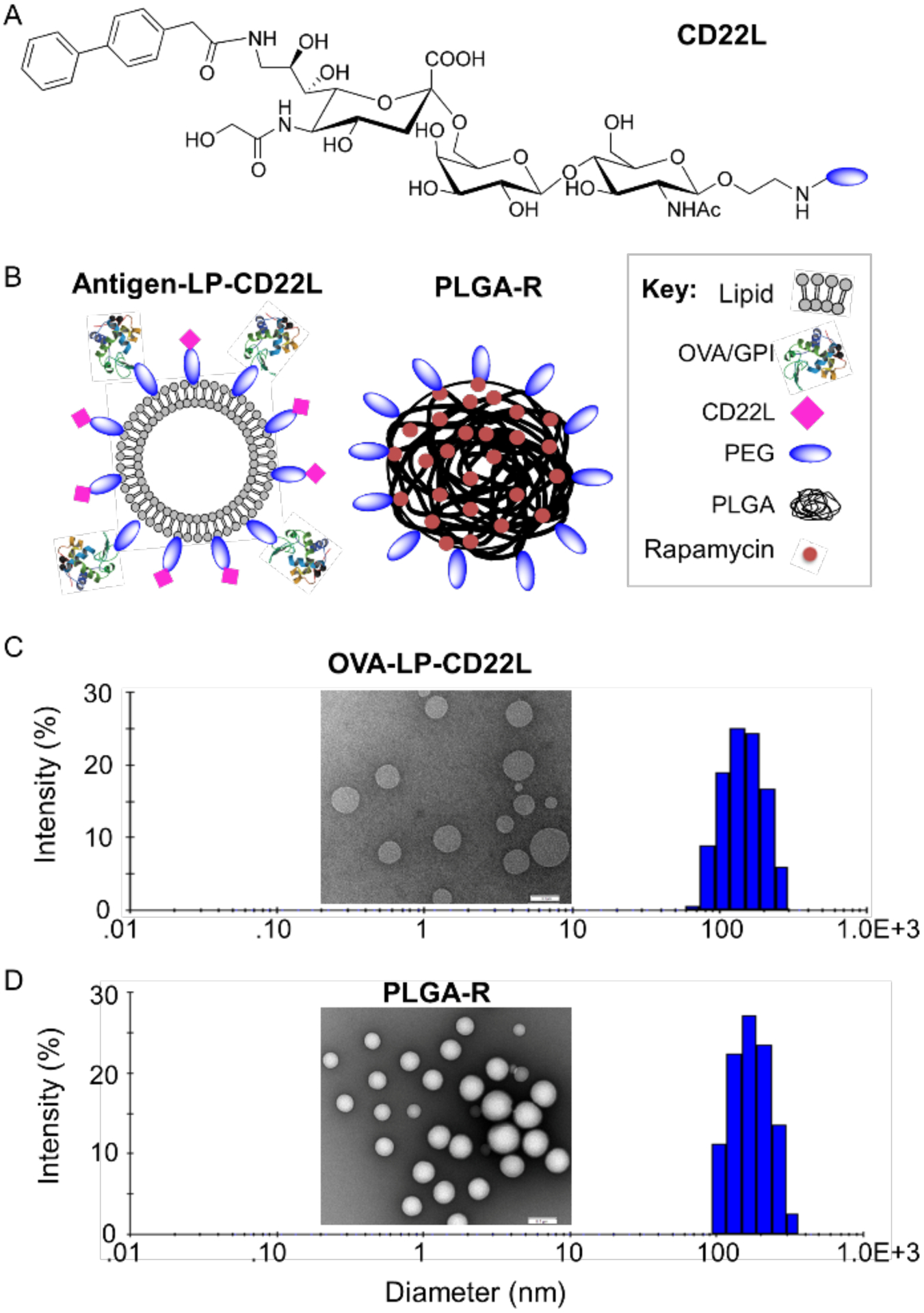
Nanoparticle formulation and characterization. (A) Chemical structure of the murine CD22 ligand linked to PEG. (B) Schematic illustration of targeted OVA/GPI-STALs (OVA/GPI-LP-CD22L) and PLGA rapamycin (PLGA-R) nanoparticles. (C-D) Representative transmission electron microscopic images of OVALP-CD22L (inset C) and PLGA-R (inset D), and hydrodynamic size distribution of OVA-LP-CD22L (156±32; plot C) and PLGA-R (182±31; plot D) as determined by dynamic light scattering.