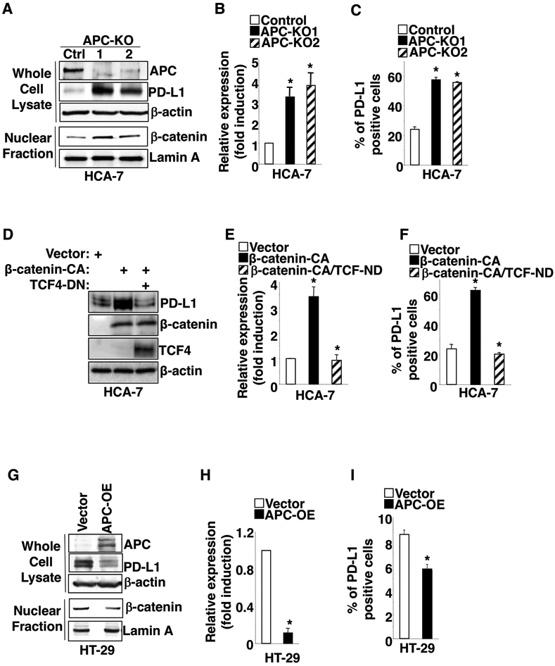
Fig. 2. Knockout or overexpression of APC alters PD-L1 expression via a β-catenin/TCF4 pathway.

(A-C) APC gene was deleted by CRISPR/Cas9 in HCA-7 cells. The protein levels of indicated genes in whole cell lysate or nucleus were measured by Western blot (A), whereas PD-L1 levels at mRNA and cell surface were measured by q-PCR (B) and flow cytometry (C) in HCA-7/control, HCA-7/APC-KO1, and HCA-7/APC-KO1 cells. APC-KO: APC knockout. (D-F) The protein levels of indicated genes in whole cell lysate were measured by Western blot (D), whereas PD-L1 levels at mRNA and cell surface were measured by q-PCR (E) and flow cytometry (F) in HCA-7/vector, HCA-7/β-catenin-CA, and HCA-7/β-catenin-CA+TCF-DN cells. (G-I) The protein levels of indicated genes in whole cell lysate and nucleus were measured by Western blot (G), whereas PD-L1 levels at mRNA and cell surface were measured by q-PCR (H) and flow cytometry (I) in HT-29/vector and HT-29/APC-OE cells. APC-OE: APC overexpression. Each Western blot image is representative of three independent experiments with similar results. Data are presented as mean ± SEM of three independent experiments. *p<0.05.
