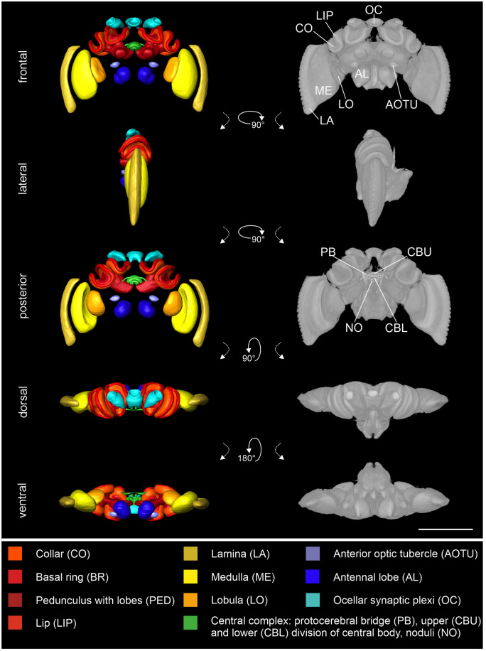
Fig. 2.
Standard brain atlas of Bombus terrestris. Left column: Shape-based average of surface reconstruction from a frontal, lateral, posterior, dorsal, and ventral perspective. The color code at the bottom represents the colors of the reconstructed neuropils. Right column: Direct volume rendering of averaged raw data from a frontal, lateral, posterior, dorsal, and ventral perspective. For better visibility of the neuropils in the central brain, the remaining neuropils (RN) were excluded in this figure. The whole standard including the RN is provided in the supplementary information (Supplementary Fig. S2). Scale bar = 1000 µm

