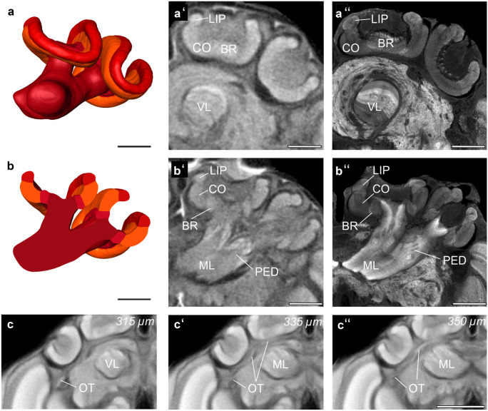
Fig. 5.
Mushroom bodies. (a) Shape-based average of surface reconstruction of the mushroom body (MB) with pedunculus (PED), vertical lobe (VL), medial lobe (ML), and the calyx (basal ring: BR, collar: CO, lip: LIP). (b) Virtual section through 3D reconstruction shown in (a). (a′ and b′) Anterior view of micro-CT scans at two different levels showing the compartments of the MB. (a′) Anterior slice shows the BR, CO, LIP, and VL. (b′) Posterior slice shows the BR, CO, LIP, and PED. (a″ and b″) Confocal image (frontal optical slice) of the MB, stained with an antiserum against synapsin. Optical section levels correspond to those in (a′) and (b′). (c, c′, and c″) The optic tracts (OT) consisting of the anterior superior optic tract (ASOT), the anterior inferior optic tract (AIOT), and the lobula optic tract (LOT). The tracts are shown here in different depths of the standard brain micro-CT data. Since they run in parallel, they cannot be distinguished here. Scale bars = 200 µm (a-b″), 500 µm (c-c″)

