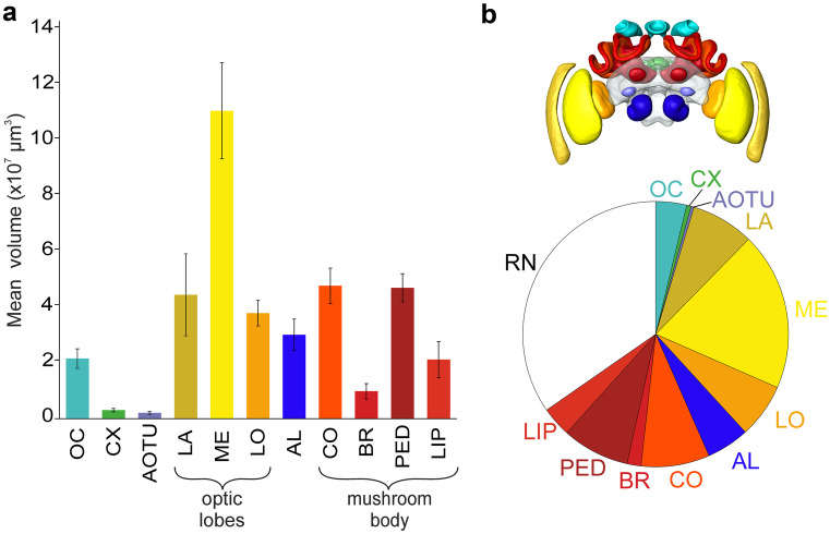
Fig. 9.
Volumetric analysis of the bumblebee brain. (a) Mean volume and standard deviation of the different neuropils in the ten individual bumblebee brains. For better visibility of the smaller volumes the remaining neuropils were left out here. (b) Upper illustration shows the shape-based average of reconstructed neuropils. The lower figure highlights the proportion of the reconstructed neuropils. Neuropils: antennal lobes (AL), anterior optic tubercle (AOTU), basal ring (BR), central complex (CX), collar (CO), lamina (LA), lip (LIP), lobula (LO), medulla (ME), ocellar synaptic plexi (OC), peduncle (PED), and remaining neuropils (RN)

