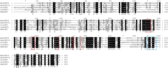
FIGURE 4.
Multiple sequences alignment of MaASMTs with the ASMTs from other plant species. The positions of the different conserved domains are represented by different colored boxes. The conserved motif for the S-adenosine-L-methionine binding is boxed in red, putative substrate-binding residues are boxed in black, and catalytic residues are boxed in blue.

