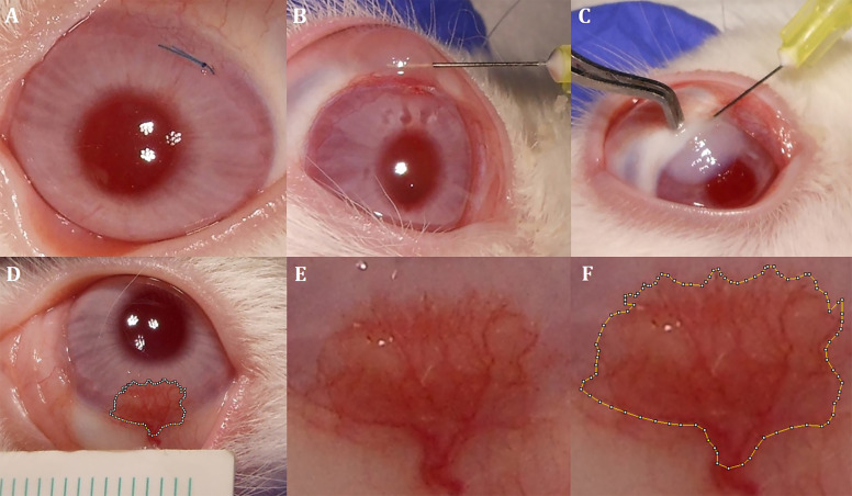
Fig. 1. Interventions to the cornea and measurement of CoNV area.
A 8/0 silk suture placement on the superior part of the cornea. B Subconjunctival administration technique. C Intrastromal administration technique. D Standard imaging of CoNV area and adjacent ruler. E Magnified CoNV area without marking. F Magnified CoNV area surrounded by manual marking.

