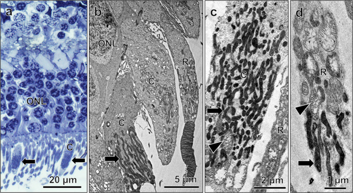
Fig. 2. Mitochondrial status in human photoreceptor cells.
Light micrograph (a) and electron micrographs (b–d), showing mitochondrial features in photoreceptor cells. (a) Dark mitochondria (arrows) in cones (C) are visible in semithin section. b–d Show dark, condensed mitochondria in cone inner segments (arrows) and rod (R, arrow in d), few swollen mitochondria are seen amongst them (c, d; arrowheads). From 83-year (a, c; nasal part) and 85-year (b, d; perifoveal part)-old donor retina.

