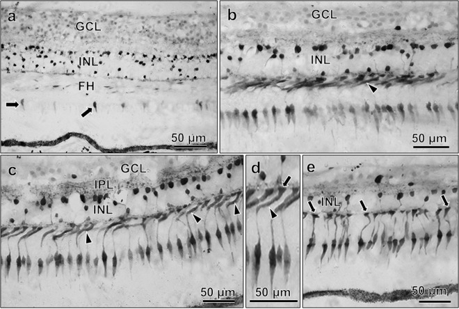
Fig. 3. Calbindin D immunoreactivity in 94-year-old donor retina.
In the parafovea (a), immunoreactivity is weakly present in few cone inner segments (arrows). It is strong in many cones of the perifovea (b, c), where abnormal axonal swellings (arrowheads) before terminating into pedicles are seen, as shown in enlarged view in d; arrow denotes pedicle (in d). In mid-peripheral cones, the axons terminate straight into pedicles (arrows, e). In all areas, the INL shows strong immunoreactivity.

