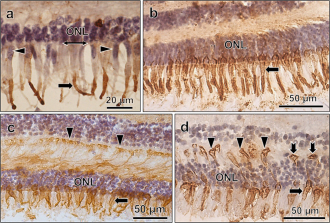
Fig. 4. Pattern of L/M opsin immunoreactivity in aging cones.
L/M opsin immunoreactivity in cones from parafoveal (a) and mid-peripheral retina (b–d). In (a), immunoreactivity is present in COS only (arrow), whereas in (b–d), it is also present in membranes of inner segments (arrows) and cone terminals (arrowheads; c, d). In (d), the cone pedicles lie at different levels. Two of them lie close to the ONL (notched arrows), indicating their retraction from the OPL. Counterstained with hematoxylin. In (a), note the prolapse of cone nuclei (arrowheads) outer to the OLM (right-left arrows). From 75-year- (a), 81-year- (b), 83-year- and (c) 85-year- (d) old retinas.

