
Fig. 7. Photoreceptor cell status in aging retina.
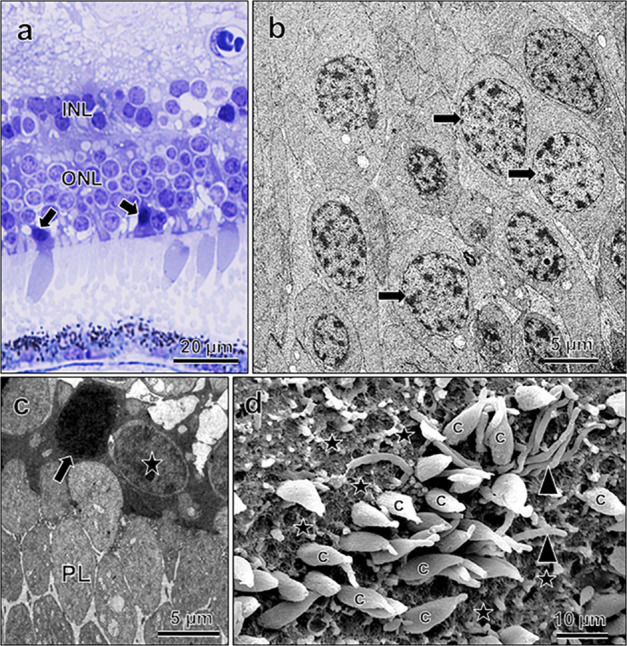
a Light micrograph showing dark, condensed cone nuclei (arrows). Electron micrographs of normal, uncondensed nuclei (b, arrows) and one condensed photoreceptor cell nucleus (c, arrow), note one normal nucleus adjacent to it (star). PL photoreceptor layer (in c). d Scanning electron micrograph of macular photoreceptor cell layer, showing empty spaces (stars), indicating loss of cones (c); note few rods (arrowheads) are present. From 89-year- (a, c; mid-periphery), 62-year- (c, macular), and 89-year- (d; parafoveal region) old retinas.