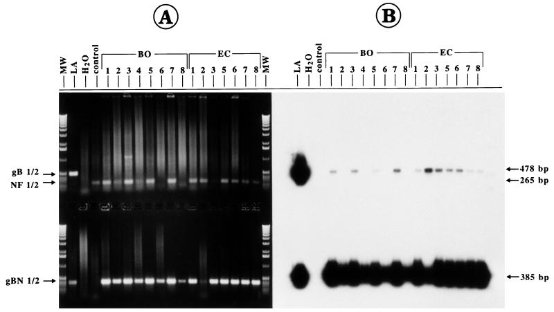
FIG. 4.
Detection of BHV-1 DNA in blood samples from field-virus-infected cattle (animals BO-1 to BO-8 and EC-1 to EC-8) after gB-1–gB-2 PCR (upper part) and gBN-1–gBN-2 PCR (lower part). As a positive control, 10 ng of strain LA DNA was used as a template (lane LA), and as negative controls, amplifications were performed with DNA derived from noninfected cattle (lane control) and in the absence of template (lane H2O). Lane MW, separation of molecular size markers (1-kbp ladder). The control NF1-NF2 PCR (265 bp) was performed separately, but the PCR products were separated together with the gB-1–gB-2 PCR products in the same gel slots. (A) The PCR products were analyzed by electrophoresis in an ethidium bromide-stained agarose gel (1.0%). No visible gB1/2 product (478 bp) was amplified by the first PCR, although in most cases a successful PCR could be demonstrated by a positive NF1-NF2 amplification (upper panel). Nested PCR (gBN-1–gBN-2; 385 bp) showed the presence of BHV-1 DNA in blood samples from all animals in herds BO and EC (lower part). (B) Southern blot hybridization of the gel shown in panel A with the radioactively labelled internal probe already revealed specific amplification in some of the blood specimens after the first PCR (upper part) and corroborated the positive results of the nested PCR (lower part).

