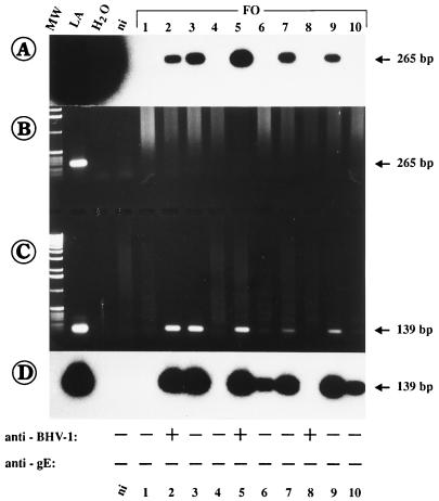
FIG. 6.
Detection of BHV-1 DNA in blood samples (herd FO) by gE-1–gE-2 (A and B) and nested gEN-1–gEN-2 PCR (C and D). Lane ni, the result obtained with blood from noninfected cattle. The sizes of the PCR products are indicated to the right. The ELISA results for the detection of serum antibodies against BHV-1 and against gE of BHV-1 are given at the bottom for each animal. As can be seen, samples 2, 3, 5, 6, 7, 9, and 10 were clearly positive after nested gEN-1–gEN-2 PCR, but the corresponding serum samples were all negative by gE-specific ELISA.

