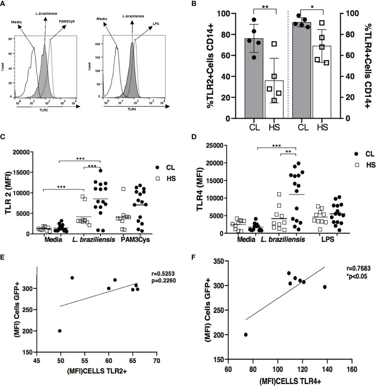
Figure 1.
TLR2 and TLR4 expression in L. braziliensis-infected monocytes from cutaneous leishmaniasis patients and healthy subjects. Monocytes in PBMC from CL patients (n = 15) and HS (n = 10) were infected with L. braziliensis (5: 1) for 2 hours and then the cells were labeled with anti-CD14 antibodies for the characterization of monocytes. Figure (A) shows the representative histogram from TLRS expression in CL patients. Frequency of TLR2 and TLR4 receptors in in the same CD14+ cells is showed in Figure (B) MFI of TLR2 and TLR4 in CD14+ infected cells are presented in figure (C) and (D) As a positive control of infection, Pam3Cys (TLR2 agonist) and LPS (TLR4 agonist) were used. Receptor expression was performed by flow cytometry. All p values were obtained using the Mann Whitney and Wilcoxon signed-rank test *p < 0.05, **p < 0.01 and ***p < 0.001. The Pearson correlation analyses was performed between GFP MFI and TLR2 (E) and TLR4 (F).

