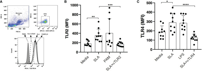
Figure 5.
Expression of TLR2 and TLR4 after stimulation with leishmania soluble antigen (SLA). PBMCs from CL patients (n=10) were stimulated with 5ug/ml SLA for 2 hours. Pam3Cys (a TLR2 agonist) and LPS (a TLR4 agonist) were used as positive controls (A).The expression of TLR2 (B) and TLR4 (C) were quantified by flow cytometry. Data are representative of median mean fluorescence intensity (MFI) values. Wilcoxon signed-rank test; *p<0.05, **p<0.01, ***p<0.001.

