Figure 1.

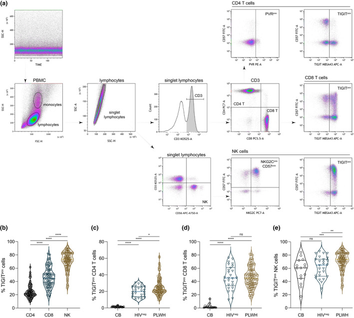
TIGIT expression on immune cell subsets from cord blood and peripheral blood of PLWH and HIV‐seronegative adults. (a) Quality of flow cytometry data acquisition was monitored by side scatter over time. Lymphocytes were identified by scatter characteristics and doublet exclusion. T cells were identified as CD3+ lymphocytes and distinguished by either CD4 or CD8 expression, and NK cells were CD3‐CD56+ lymphocytes. Subsets of CD4+ T cells, CD8+ T cells and NK cells were further demarcated by TIGIT and CD57 expression, and CD4+ T cells were analysed for PVR expression and NK cells for NKG2C and CD57 expression. (b) Compiled TIGIT expression levels on CD4+ T cells, CD8+ T cells and NK cells from PLWH (n = 95). Friedman test ****P < 0.0001. Expression of TIGIT on (c) CD4+ T cells, (d) CD8+ T cells and (e) NK cells from nascent (CB, n = 15), HIV‐seronegative (HIVneg, n = 26) and PLWH (n = 95) was compared. Kruskal–Wallis test *P = 0.0408 **P = 0.0082 ***P = 0.0008 ****P < 0.0001. Horizontal lines bisecting groups represent median with IQR.
