Figure 2.
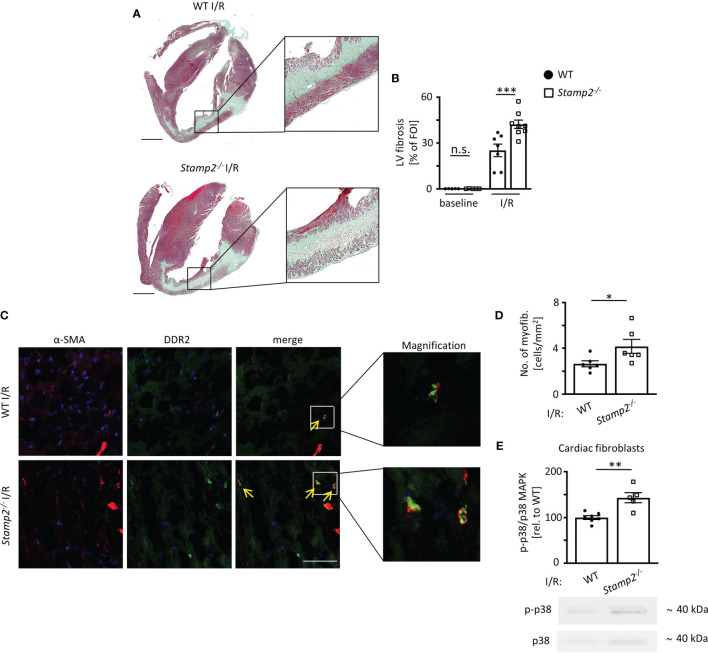
Stamp2 deficiency aggravates I/R-induced LV fibrotic remodeling. (A) Representative images of Masson’s trichrome-stained cardiac sections of WT and Stamp2-/-animals 7 days after I/R. Scale bar=1mm. (B) Quantification of left ventricular fibrotic areas stained in green (n=5/5/7/8). (C) Representative immunofluorescence stainings imaged by confocal microscopy for the myofibroblast marker α-SMA (α-smooth muscle actin; red), for the fibroblast marker DDR-2(discoidin domain-containing receptor 2; green) and for nuclei (DAPI, blue). Scale bar=50μm. (D) Quantitative analaysis of myofibroblasts within the peri-infarct region (n=6/6). (E) Relative phosphorylation of p38 MAPK (p-p38/p38 MAPK) in isolated primary fibroblasts from hearts 3 days post I/R (n=7/5). Graphs show Mean ± SEM. Full blots are shown in Supplemental Figure S4 . Significance was determined by two-way ANOVA followed by Tukey post-hoc test (B) or by unpaired Student’s t-test (D, E). *P < 0.05, **P < 0.01, ***P < 0.001. n.s., not significant.

