Figure 2.
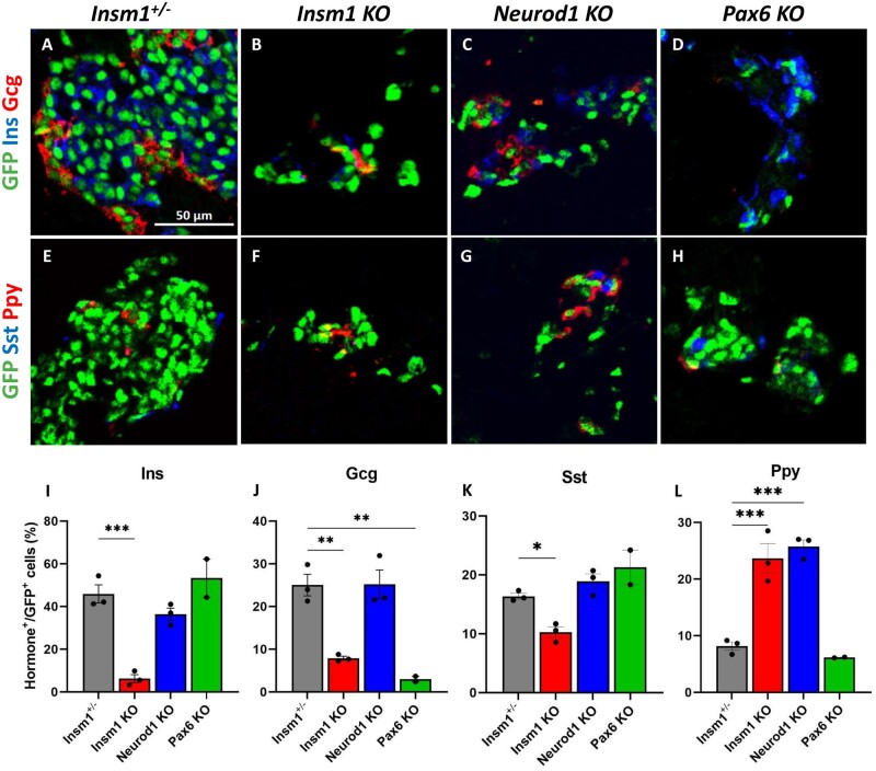
Impaired differentiation of pancreatic endocrine cells in Insm1, Neurod1, and Pax6 KO embryos. (A–D) Immunofluorescence labeling of pancreata from E18.5 Insm1GFP-expressing embryos using antibodies against GFP (green) that marks pre-endocrine cells, and pancreatic hormones insulin (Ins, blue), and glucagon (Gcg, red). Compared with Insm1+/− mice (A), mice lacking Insm1 (B), Neurod1 (C), and Pax6 (D) exhibit a decrease in total number of endocrine cells, altered endocrine cell morphology and numbers of hormone expressing cells. (E–H) Immunofluorescence labeling of pancreata from E18.5 embryos using antibodies against GFP (green), pancreatic polypeptide (Ppy, red), and somatostatin (Sst, blue) shows altered numbers of hormone expressing cells in Insm1 (F), Neurod1 (G), and Pax6 (H) KO mice. (I–L) Quantification of a percentage of hormone-positive cells among GFP-positive endocrine cells demonstrates defects in differentiation of cells positive for hormones: insulin (Ins) (I), glucagon (Gcg) (J), somatostatin (Sst) (K), and pancreatic polypeptide (Ppy) (L) in Insm1, Neurod1, and Pax6 KO embryonic pancreata in comparison with Insm1+/−. Error bars indicate SEM (n = 3); P-values were determined by one-way ANOVA test. Asterisks indicate P-values of *<0.05, **<0.01, and ***<0.001. Scale bars: 50 μm.

