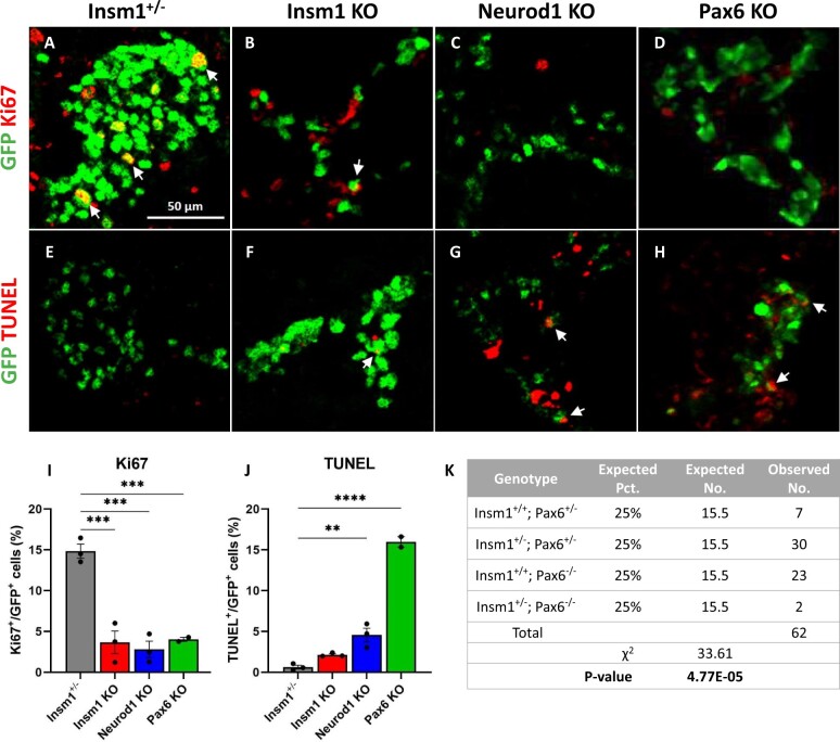
Figure 3.
Decreased proliferation of endocrine cells in Insm1, Neurod1, and Pax6 KOs, and increased apoptosis in Neurod1 and Pax6 KO embryos. (A–D) Immunofluorescence labeling of pancreatic tissues from E18.5 Insm1GFP-expressing embryos using antibodies against GFP (green) that marks all pre-endocrine cells, and antibodies against cell proliferation marker Ki67 (red). Compared to controls (A), mice lacking Insm1 (B), Neurod1 (C), and Pax6 (D) exhibit a decrease in the number of Ki67-positive endocrine cells. Arrows indicate cells co-expressing GFP and Ki67. (E–H) Immunofluorescence labeling of pancreatic tissues from E18.5 embryos with antibodies against GFP (green) and TUNEL assay (red) marking positive apoptotic events. Compared with Insm1+/− mice (E), mice lacking Insm1 (F), Neurod1 (G), and Pax6 (H) exhibit an increase in the number of endocrine cells positive for both TUNEL and GFP. Arrows indicate TUNEL positive cells co-expressing GFP. (I) Quantification of a percentage of Ki67-positive cells among GFP-positive cells demonstrates a proliferation defect in endocrine cells from Insm1, Neurod1, and Pax6 KO pancreata at E18.5. (J) Quantification of a percentage of TUNEL-positive cells among GFP-positive cells demonstrates increased apoptosis in endocrine cells in Neurod1 and Pax6 KO pancreata at E18.5. Error bars indicate SEM (n = 3); P-values were determined by one-way ANOVA test. Asterisks indicate P-values of *<0.05, **<0.01, and ***<0.001. Scale bars: 50 μm.

