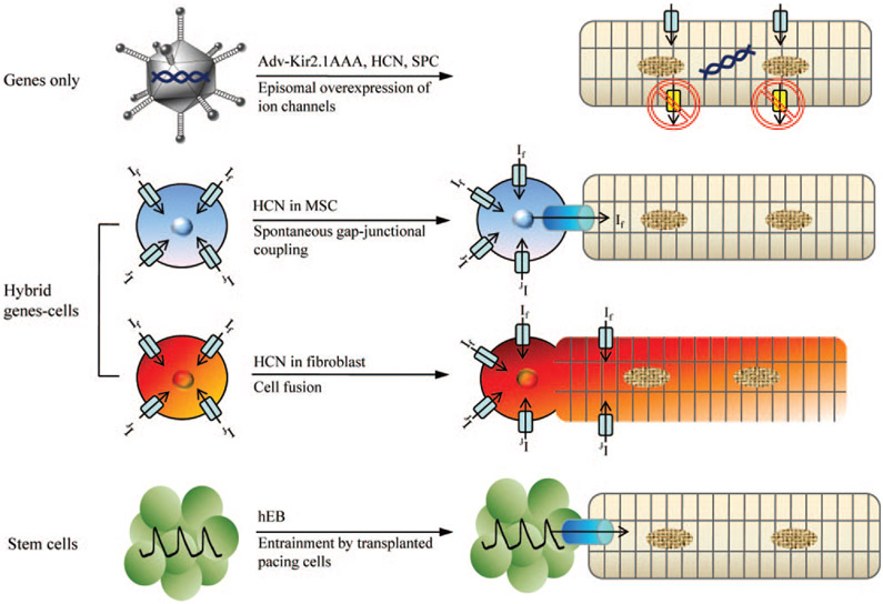
Figure 6.
A summary of different approaches to creating a biological pacemaker. First approach (top row) is a strict gene therapy in which Kir2.1AAA, HCN, or synthetic pacemaker channel genes are overexpressed in myocytes via adenoviral delivery. Kir2.1 dominant negative proteins suppress repolarizing, outward currents whereas pacemaker channels directly contribute to diastolic membrane potential depolarization. Delivering If by MSCs requires gap-junctional coupling between myocytes and MSCs (second row). In the cell fusion approach (third row), If and the pacemaker activity arise from the HCN channels expressed on the cell membrane of the heterokaryon, without the need for gap-junctional coupling. Spontaneously beating human EBs and cardiospheres transduce their pacemaker activity to cardiomyocytes via electrotonic cell–cell coupling (fourth row).

