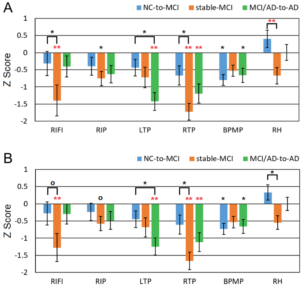
Fig. 1.

Z scores of normalized longitudinal rCBF changes in the NC-to-MCI, stable-MCI, and MCI/AD-to-AD subjects relative to stable-NC subjects: (A) without PVE correction and (B) with PVE correction. The significance level is represented by * (0.01 ≤ p ≤ 0.05) and ** (p < 0.01). Red stars (*) represent the significant differences after FWE correction. RIFI: right inferior frontal and insular, RIP: right inferior parietal, LTP: left temporoparietal, RTP: right temporoparietal, BPMP: bilateral posterior and middle cingulate and parietal, and RH: right hippocampus.
