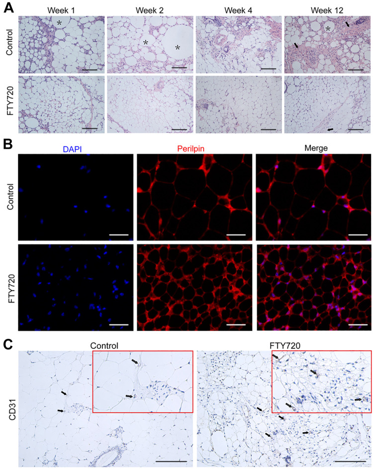
Figure 2.
FTY720 improved fat graft structure integrity in vivo. (A) The histological changes of the fat graft at week 12 were detected by HE staining. Oil cysts and fibrosis structures were indicated by black asterisks and black arrows, respectively. Scale bars = 120 µm. (B) Perilipin immunostaining of the fat graft at week 12 was used to stain the mature adipocytes. Scale bars = 60 µm. (C) Representative IHC staining images of CD31 in the FTY720 group and control group at week 12. Scale bars = 120 µm.

