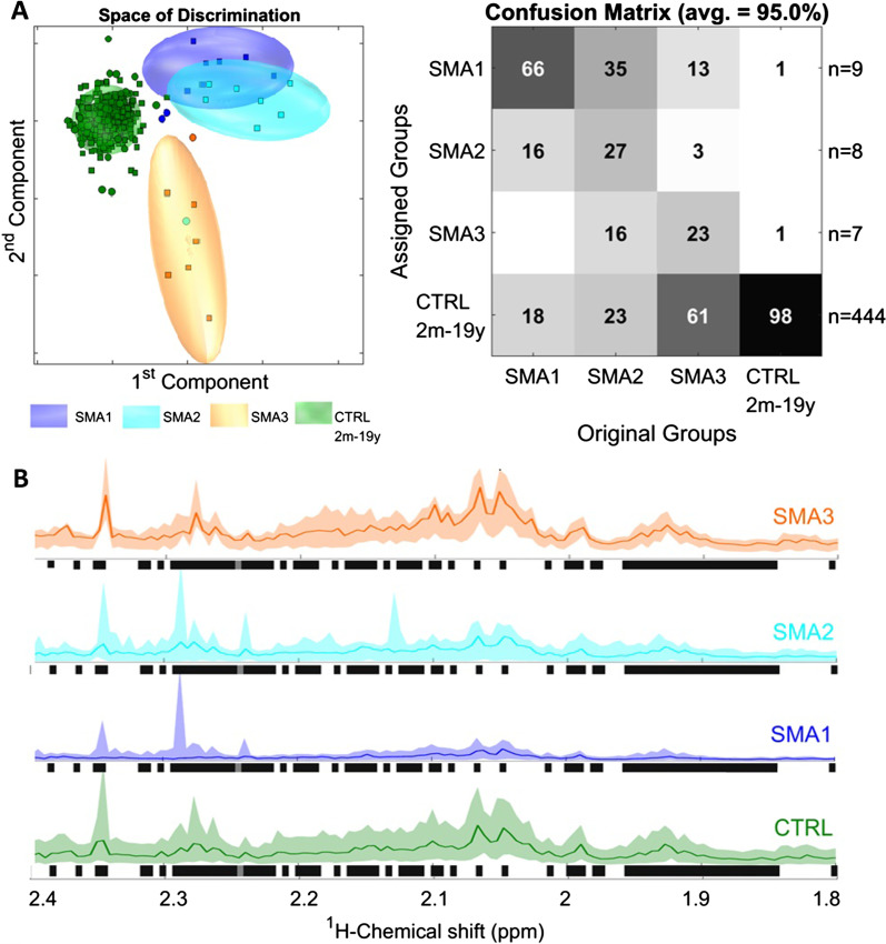
Fig. 4.
Disease prediction. A PCA/CA classification and MCCV of the SMA 1, 2 and 3 group and an age-matched healthy control group showed clear discrimination between the SMA 1 and SMA 3 group with some overlap between the SMA 1 and 2 group as well as the SMA 3 group and controls. PCA/CA was performed on 1,000 variables from 0.5 to 10 ppm (exclusion: see Materials and Methods) with Expl. Variance of 99.9%. The Confusion Matrix is the result of 100 Monte-Carlo-Runs (MC) with sevenfold CrossValidation (CV). Space of discrimination is one representation of the modelling samples in 2-dimensions. B Expansion of 6% of overall 1H NMR spectra. The Kruskal–Wallis test of 1H NMR spectra of the different SMA types and controls revealed significant protein backgrounds in the aliphatic region as one of the major discriminants. Colored lines represent medians and colored areas correspond to variation (12.5–87.5% quantile) of 1H-NMR spectra. Spectral regions highlighted in black illustrate significant differences between the 4 groups (p-value < 0.01). SMA: spinal muscular atrophy. CTRL: control

