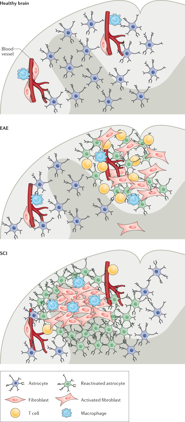
Fig. 3. Organization of the glial and fibrotic scars.
In a healthy spinal cord, perivascular fibroblasts and macrophages reside in perivascular spaces1,9. In experimental autoimmune encephalomyelitis (EAE), a model of multiple sclerosis, neuroinflammatory lesions form in the white matter. These lesions include infiltrating immune cells, such as T cells, and a scar consisting of nearby fibroblasts and reactive astrocytes4,104. In spinal cord injury (SCI), a scar also forms in the area of the injury. However, the core of the injury site consists of an inner fibrotic scar containing extracellular matrix proteins, activated fibroblasts, microglia, and macrophages and an outer glial scar consisting of reactive astrocytes26,79,93.

