. 2021 Sep 20;12(40):13379–13391. doi: 10.1039/d1sc04338b

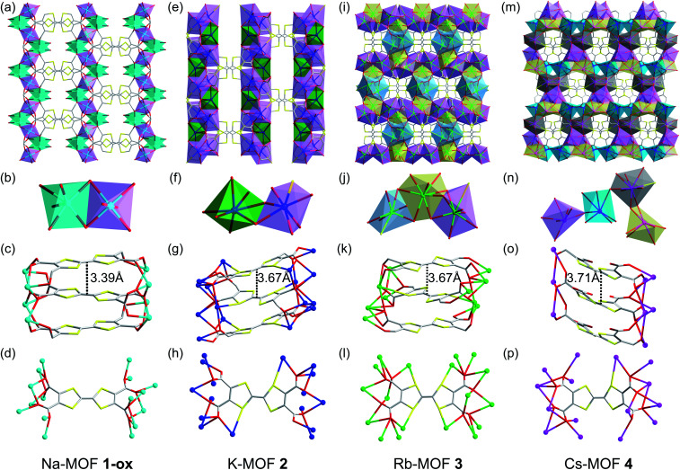
Fig. 2. (a–d) Na-MOF (1-ox): (a) the crystal structure viewed along the c-axis, (b) the SBU contains two distinct hexacoordinated Na+ ions bridged by carboxylate groups, (c) the alignment of perfectly planar TTFTC ligands π-stacked along the c-axis, and (d) the coordination pattern of each TTFTC ligand. (e–h) K-MOF (2): (e) the crystal structure viewed along the c-axis, (f) the SBU features two distinct hepta-coordinated K+ ions bridged by the carboxylate groups, (g) the alignment of slightly bent TTFTC ligands π-stacked along the c-axis, and (h) the coordination pattern of each TTFTC ligand. (i–l) Rb-MOF (3): (i) the crystal structure viewed along the c-axis, (j) the SBU contains two octa-coordinated (yellow and violet polyhedrons) and one hepta-coordinated (blue polyhedron) Rb+ ions, (k) the alignment of puckered TTFTC ligands π-stacked along the c-axis, and (l) the coordination pattern of each TTFTC ligand. (m–p) Cs-MOF (4): (m) the crystal structure viewed along the a-axis, (n) the SBU contains four distinct Cs+ ions (cyan, grey, violet, and yellow polyhedrons) bridged by the carboxylate groups, (o) the alignment of boat-shaped TTFTC ligands π-stacked along the b-axis, and (p) the coordination pattern of each TTFTC ligand. C: grey, O: red, S: yellow, Na+: cyan ball, K+: navy blue ball, Rb+: green ball, and Cs+: pink ball. The asymmetric units and additional structural features of all four MOFs are shown in Fig. S1..

Fig. 2