. 2021 Aug 3;12(40):13292–13300. doi: 10.1039/d1sc01560e

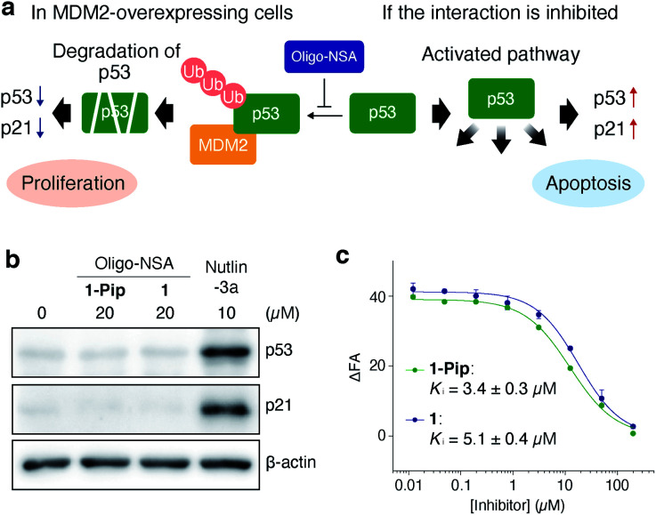
Fig. 2. Initial design of oligo-NSA-based inhibitors of MDM2–p53 interaction. (a) Schematic illustration of the negative regulation of p53 function by MDM2 and activation of the p53 pathway via the inhibition of MDM2–p53 interaction. (b) Intracellular protein levels of p53, p21, and β-actin. SJSA-1 cells were treated with 20 μM oligo-NSA for 8 h and cell lysates were analyzed by western blotting. Lysate of SJSA-1 cells treated with a known inhibitor of the MDM2–p53 interaction, Nutlin-3a, was analyzed as a positive control. Representative results of three independent experiments are shown. Other results are shown in Fig. S1. (c) Inhibitory curves of 1-Pip and 1 against the interaction between fluorescently labeled PMI peptide and MDM2 from a competitive FP assay. Y-axis indicates change in fluorescence anisotropy (ΔFA). Error bars represent standard deviations of triplicates.

Fig. 2