. 2021 Sep 9;12(40):13434–13441. doi: 10.1039/d1sc00193k

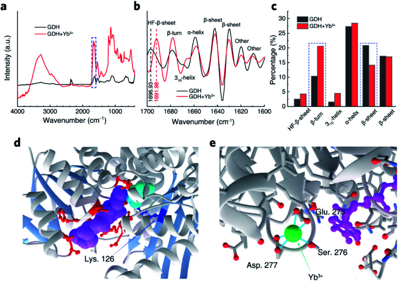
Fig. 6. Regional changes of GDH structure. (a) ATR-FTIR spectra of GDH pre-incubated with or without Yb3+. Dashed frame denotes the amide I band region. (b) Fourth derivation of amide I band. (c) Compositional percentages of different GDH secondary structures by integrating peak areas of deconvoluted bands. (d) Zoomed image of GDH active site with bound NADH (purple), glutamate (cyan) and residues binding NADH (red). Lys. 126 attacking glutamate intermediate and β-sheets guarding the active site are highlighted in dark blue and light blue, respectively. (e) The most plausible anchoring site for Yb3+ facing three potential ligand residues, Glu. 275, Ser. 276 and Asp. 277, which constitute a β-turn near bound NADH.

Fig. 6