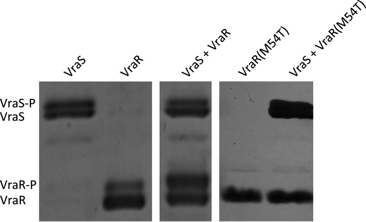
FIG 1.
Phos-tag SDS-PAGE of in vitro phosphorylation of VraS, VraR, and VraR(M54T) after incubation with ATP. Phosphorylated bands (P) are indicated. The figure shows two representative gels. The presence of a seemingly phosphorylated protein band in the VraR control in the absence of VraS indicates either a partial phosphorylation of VraR by a kinase from E. coli or an impurity of the VraR protein preparation.

