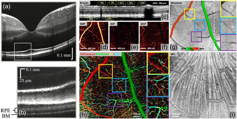
Fig. 8.
(a)–(b) In vivo human retinal visible light OCT; (b) inset from (a). Adapted from Ref. 274. (c) B-scan image of a brown Norway rat retina using visible-light OCT. NFL, nerve fibre layer; GCL, ganglion cell layer; IPL, inner plexiform layer; INL, inner nuclear layer; OPL, outer plexiform layer; and BM, Bruch’s membrane. (d)–(f) En face images of vascular/capillary plexuses. SVP projected in the NFL and GCL slabs. ICP projected in the slab containing the inner border of the INL. DCP projected in the slab containing the outer border of the INL. (g) En face structural image projected from the ILM to BM, overlaid with measured oxygen saturation () values in major vessels to differentiate arteries from veins in an animal breathing 100% . Interplexus capillaries (white arrows) appear as dark spots due to greater light absorption than neighbouring capillaries. (h) Overlaid en face angiograms of three vascular/capillary plexuses to demonstrate the detailed organization of the retinal circulation. Examples of interplexus capillaries (indicated by white arrows in the enlarged images) were validated by observing their presence in corresponding locations. (i) En face projection of the NFL slab. The SVP was found to run anterior to the nerve fibre bundles (bright radial striations), which appear posterior to the vessels. The interplexus capillaries (black arrows) penetrate between NFL bundles and connect the SVP to the ICP and DCP. (c)–(i) Adapted from Ref. 276.

