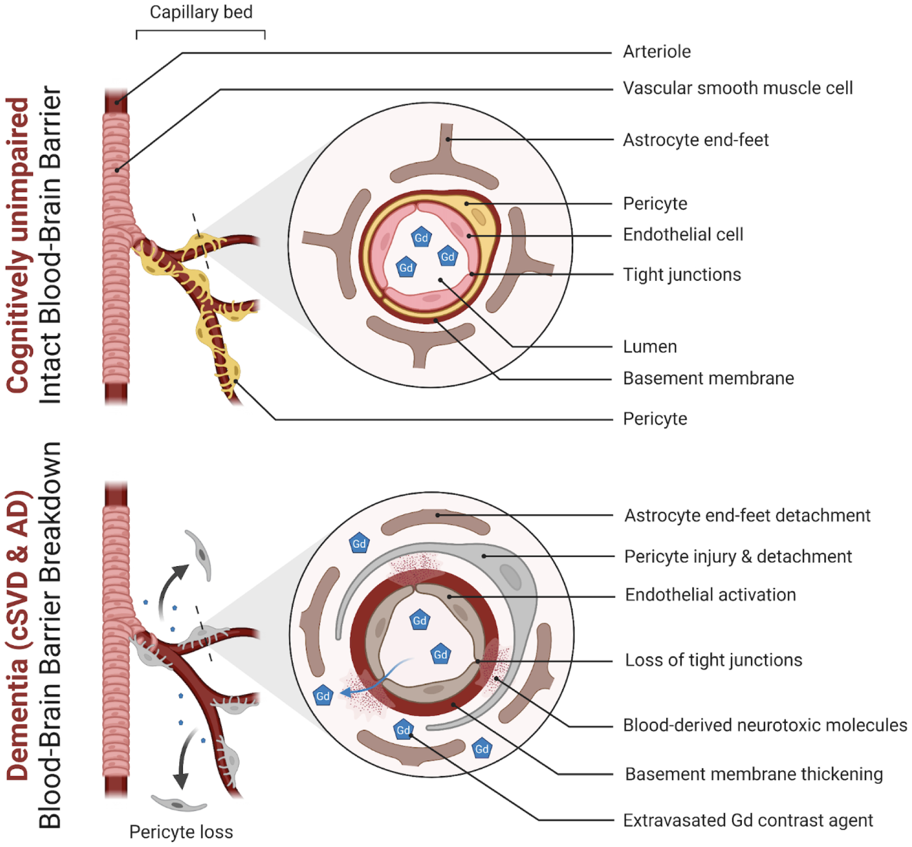
Figure 1. Blood-brain barrier in health and dementia.

A simplified neurovascular unit (NVU) diagram showing a healthy blood-brain barrier (BBB) with the interactive cellular network at the level of brain capillaries that comprises endothelial cells, pericytes, basal membrane, and astrocyte end-feet (top panel). In dementia, changes to endothelial cells and pericytes lead to loss of function and BBB breakdown with loss of tight junction proteins (bottom panel). Subtle extravasation of Gadolinium (Gd) contrast can be detected using dynamic contrast-enhanced magnetic resonance imaging (DCE-MRI) in both living cerebral small vessel disease (cSVD) and Alzheimer’s disease (AD) participants. Subsequent damage then occurs to the surrounding brain cells such as astrocytes, neurons, and oligodendrocytes contributing to pathology and cognitive decline (Figure created using Biorender.com).
