Figure 1.

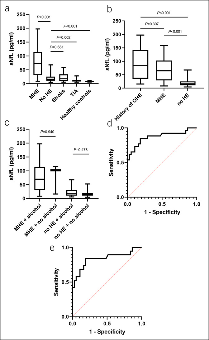
sNfL in different patient cohorts and the discriminative power of sNfL to detect MHE in patients with liver cirrhosis. sNfL in patients with liver cirrhosis with (n = 6) or without MHE (n = 38), in patients with stroke (n = 29), TIA (n = 29), and healthy individuals (n = 10) (a). sNfL in patients with liver cirrhosis and a history of OHE (n = 8), in patients with presence of MHE but no history of OHE (n = 19), and in patients without any history or presence of HE (n = 37) (b). sNfL in patients with or without alcoholic etiology of liver cirrhosis stratified by MHE status (c). Discriminative ability of sNfL to detect MHE in the total cohort of patients with liver cirrhosis (n = 64, AUC = 0.872, 95% CI 0.773–0.971; P < 0.001) (d). Discriminative ability of sNfL to detect MHE in the cohort of patients with liver cirrhosis and no history of OHE (n = 56, AUC 0.839, 95% CI 0.711–0.966; P < 0.001) (e). AUC, area under the curve; HE, hepatic encephalopathy; MHE, minimal hepatic encephalopathy; OHE, overt hepatic encephalopathy; sNfL, serum levels of neurofilament light chains; TIA, transitory ischemic attack.
