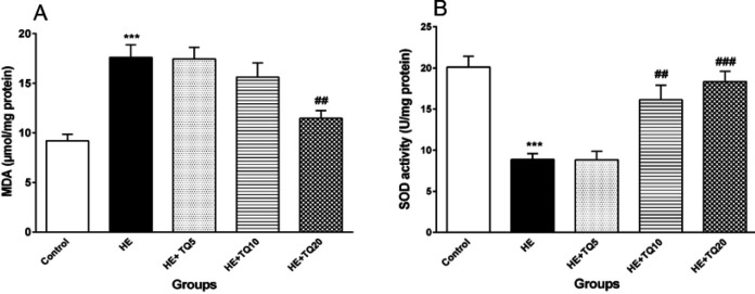
Figure 5.
Effects of TQ on the levels of malondialdehyde (MDA) in hippocampal tissue and superoxide dismutase (SOD) in TAA-induced HE rats. (A) Hippocampal tissue level of MDA; (B) levels of SOD activity in hippocampal tissue in all the groups. Data are presented as mean ± SEM (n = 5). HE: hepatic encephalopathy group, TQ: thymoquinone. *** P<0.001 vs control group, ## P<0.01 and ### P<0.001 vs HE group. Data were analyzed by one-way ANOVA followed by Tukey's post-hoc test

