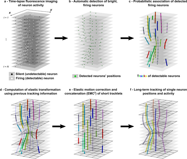
Fig 1. Multi-step EMC2 for tracking neuron activity in calcium imaging data.
a- Time-lapse imaging (N frames) of intermittent fluorescence activity of a neuron in a deforming environment (e.g. behaving animal). b- Fluorescent spots (neurons), that are significantly brighter than background, are automatically detected with a wavelet-based algorithm. c- Tracklets of detectable neurons are robustly reconstructed using probabilistic tracking algorithm (eMHT). d- Short tracklets of detectable particles are used to compute the elastic deformation of the field of view at each time frame. Associated detections in neuron tracklets are used as fiducials, and the whole deformation is interpolated using a poly-harmonic thin-plate spline function. Forward- and backward-propagated positions of tracklet particle positions are shown with a thin blue line. e- After having corrected for the deformation of the field-of-view where neurons are embedded, gaps between the end- and starting-points of tracklets are closed by minimizing the global Euclidean distance between points (dotted line). f- Finally, complete single neuron tracks over the time-lapse sequence are obtained by applying the elastic transformation of the field-of-view to concatenated tracklets.

