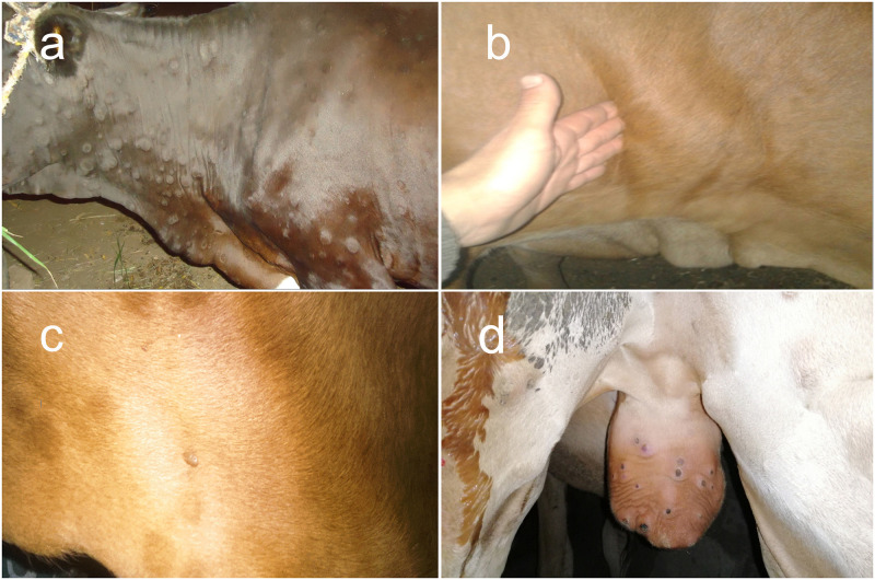
Fig 2. LSDV infected animals.
A) Cutaneous nodules covered the entire body of a LSDV infected cow. B) Enlarged and oedematous pre-scapular lymph node in a LSDV infected cow. C) LSDV infected cow infested with hard ticks. D) A 12-month-old calf with scrotal lesions. Each lesion is surrounded by a zone of necrosis (sitfast).

