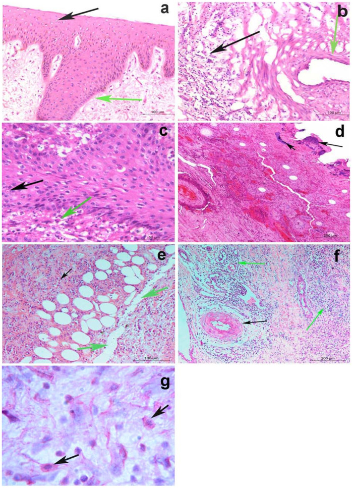
Fig 4. Histopathology of skin nodules.

a) Acanthosis (green arrow), associated with hydropic degeneration in keratinocytes of the epidermal layer of the skin (black arrow), while the next dermal layer is normal (H&E; Bar = 100μm). b) Vasculitis (green arrow), associated with massive leucocytic infiltrations in the dermal layer (black arrow) (H&E; Bar = 100μm). c) Intracytoplasmic inclusion body of LSD in the keratinocytes of the epidermal layer (black arrow), while the dermal layer shows massive leucocytic infiltrations mainly by eosinophils (green arrow) (H&E; Bar = 50μm). d) Chromatin condensation and calcium deposits in keratin layer (black arrows), while the next dermal layer shows severe hemorrhages with leucocytic aggregation (H&E; Bar = 200μm). e) Necrosis and dissociation of muscle fibers, fatty infiltrates, and leucocytic infiltration mainly by lymphocytes and few numbers of eosinophils between them (black arrows) (H&E; Bar = 100 μm). f) Dilatation of lymph vessels (LVs.), necrotizing vasculitis (black arrow), and leucocytic infiltration mainly by lymphocytes and few numbers of eosinophils between them (green arrows) (H&E; Bar = 200 μm). g) Red viral particles in macrophage of connective tissue of the dermal layer (black arrows) using alkaline phosphatase immunohistochemistry).
