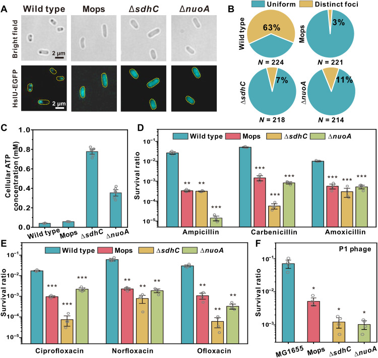
Fig. 4. Aggresome formation promotes cellular survival under fierce stresses.
(A) Bright-field and fluorescence images of MG1655, MG1655 (Mops), ΔsdhC, and ΔnuoA; (B) proportion of cells showing HslU-EGFP foci; and (C) average cellular ATP concentration in different strains, after 24 hours of culture. Cell survival rate (log scale) after 4 hours (D) β-lactam (E) fluoroquinolone, and (F) P1 phage infection. Multiplicity of infection (MOI) = 100. (Unpaired Student’s t test against wild type; error bar indicates SE; *P <0.05, **P < 0.005, and ***P < 0.0005.)

