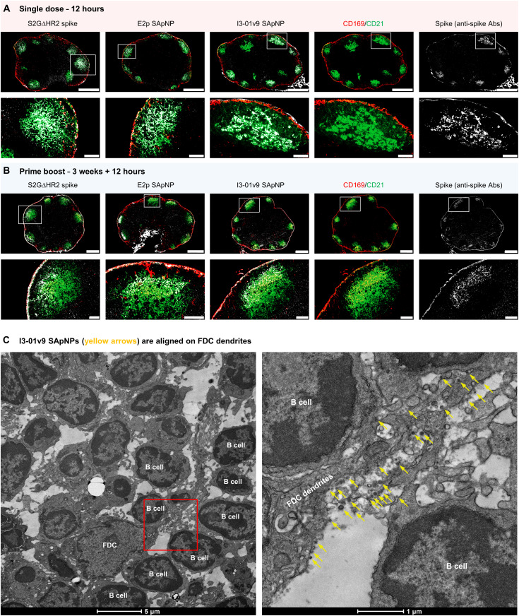
Fig. 5. SARS-CoV-2 SApNP vaccines interact with FDCs and are presented on FDC dendrites to B cells.
(A and B) S2GΔHR2 spike and S2GΔHR2-presenting E2p and I3-01v9 SApNP vaccine interaction with FDC networks in lymph node follicles 12 hours after (A) a single-dose or (B) prime-boost injections (10 μg per footpad, 40 μg per mouse). Vaccine antigens (the S2GΔHR2 spike and S2GΔHR2-presenting E2p and I3-01v9 SApNPs) colocalized with FDC networks. Immunostaining is color-coded (green, CD21; red, CD169; white, anti-spike), with scale bars of 500 and 100 μm shown for a complete lymph node and an enlarged image of a follicle, respectively. (C) Representative TEM images of an FDC surrounded by multiple B cells. S2GΔHR2-presenting I3-01v9 SApNPs (yellow arrows) presented on FDC dendrites.

