Figure 1.

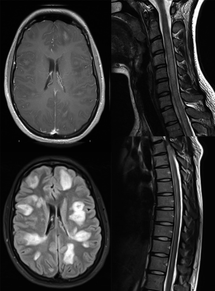
Hyperintense areas shown on MRI brain (FLAIR and post contrast T1‐weighted), cervical and thoracic spine (T2‐weighted) images.

Hyperintense areas shown on MRI brain (FLAIR and post contrast T1‐weighted), cervical and thoracic spine (T2‐weighted) images.