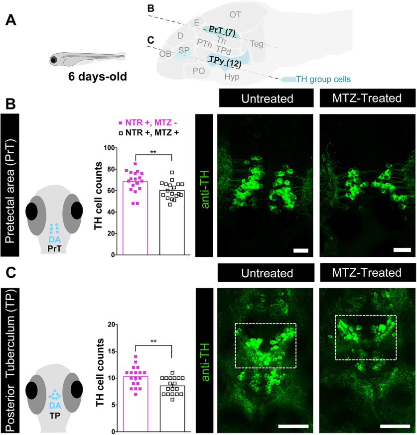
Figure 5.
Early-life OXT ablation affects dopaminergic neurons. A, top, Scheme demonstrating the position of sagittal sections through a zebrafish larva brain showing the dopaminergic clusters that were visualized by anti-TH immunofluorescence staining, and highlighting the two key dopaminergic brain areas that were affected by OXT neuronal ablation at 4–6 d old, namely, the PrT and TP. Scheme was adapted from https://zebrafishucl.org/forebrain-regions/posterior-tuberculum. B, Quantification of total PrT TH cell counts in untreated (NTR+, MTZ−) versus 4- to 6-d-old MTZ-treated (NTR+, MTZ+) larvae and respective representative images. Scale bar: 20 μm. C, Quantification of total TP TH cell counts in untreated (NTR+, MTZ−) versus 4- to 6-d-old MTZ-treated (NTR+, MTZ+) larvae and respective representative images. Scale bar: 50 μm. Data presented as mean ± SEM. Full purple squares: untreated fish (NTR+, MTZ–); open squares: MTZ-treated fish (NTR+, MTZ+); **p < 0.01. D, dorsal telencephalon; E, epiphysis; Hyp, hypothalamus; OB, olfactory bulb; OT, optic tectum; PO, preoptic; PTh, prethalamus; SP, subpallium (includes Vv, Vd, and Vs in adult); Teg, tegmentum; Th, thalamus; TPd, posterior tuberculum dorsal part; TPv, posterior tuberculum ventral part.

