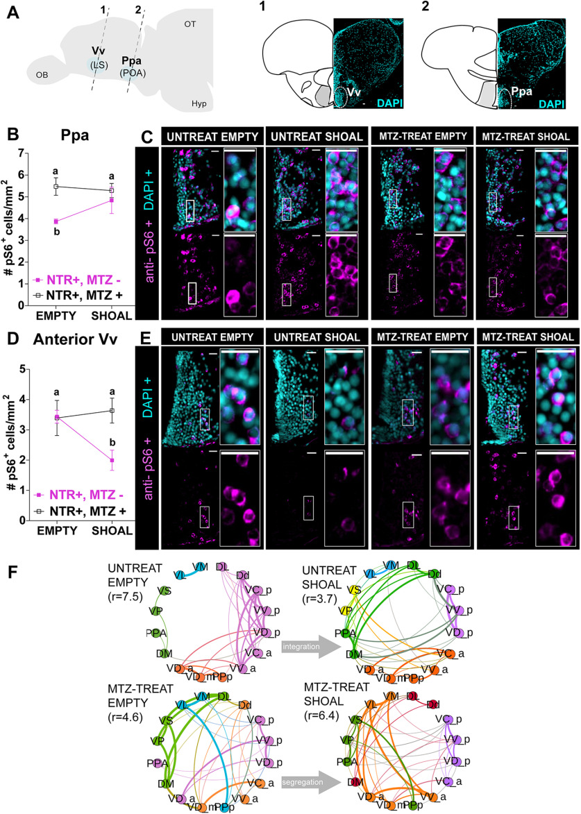
Figure 8.
Early-life OXT shapes social information processing. A, Anatomical localization of the two social responsive areas: Vv_a (1) and Ppa (2). Brain areas were identified by DAPI. B–E, Quantification of the density (cells/mm2) of cells expressing the neuronal marker pS6, visualized by anti-pS6 immunofluorescence staining, in 4- to 6-d-old MTZ-treated (open squares) or untreated adult fish (full purple squares), after exposure to either a shoal of conspecifics or an empty tank for 10 min in the Ppa area (B) and anterior Vv area (D) and respective representative examples C. E, Different letters indicate significant statistical differences (p < 0.05). F, Changes in the modular structure of functional connectivity. Modules were obtained by extracting central partition from 400 optimization of Leiden algorithm (Traag et al., 2019) on the treatment correlation matrices. Node color indicates community membership. For visualization purposes, we only show links with correlation weight >0.1. r values indicate the ratio of total edge weight within and between modules. High (low) values of r indicate more (less) segregated modular structure. Scale bar: 20 μm. Data presented as mean ± SEM. DL, lateral part of the dorsal telencephalic area; Dd, dorsal part of the dorsal telencephalic area; DM, medial part of the dorsal telencephalic area; MTZ-TREAT, treated with MTZ; UNTREAT, untreated; VC_a, central nucleus of the ventral telencephalic area (anterior); VC_p, central nucleus of the ventral telencephalic area (posterior); VD_a, dorsal nucleus of the ventral telencephalic area (anterior); VD_m, dorsal nucleus of the ventral telencephalic area (medial); VD_p, dorsal nucleus of the ventral telencephalic area (posterior); VL, ventrolateral thalamic nucleus; VM, ventromedial thalamic nucleus; VP, postcommissural nucleus of the ventral telencephalic area; VS, supracommissural nucleus of the ventral telencephalic area; VV_a, ventral nucleus of the ventral telencephalic area (anterior); VV_p, ventral nucleus of the ventral telencephalic area (posterior) DL, dorsal telencephalic area (lateral); Dd, dorsal telencephalic area (dorsal); DM, dorsal telencephalic area (medial); PPa, parvocellular preoptic nucleus (anterior); PPp, parvocellular preoptic nucleus (posterior). See Extended Data Figures 8-1, 8-2, 8-3, 8-4.

