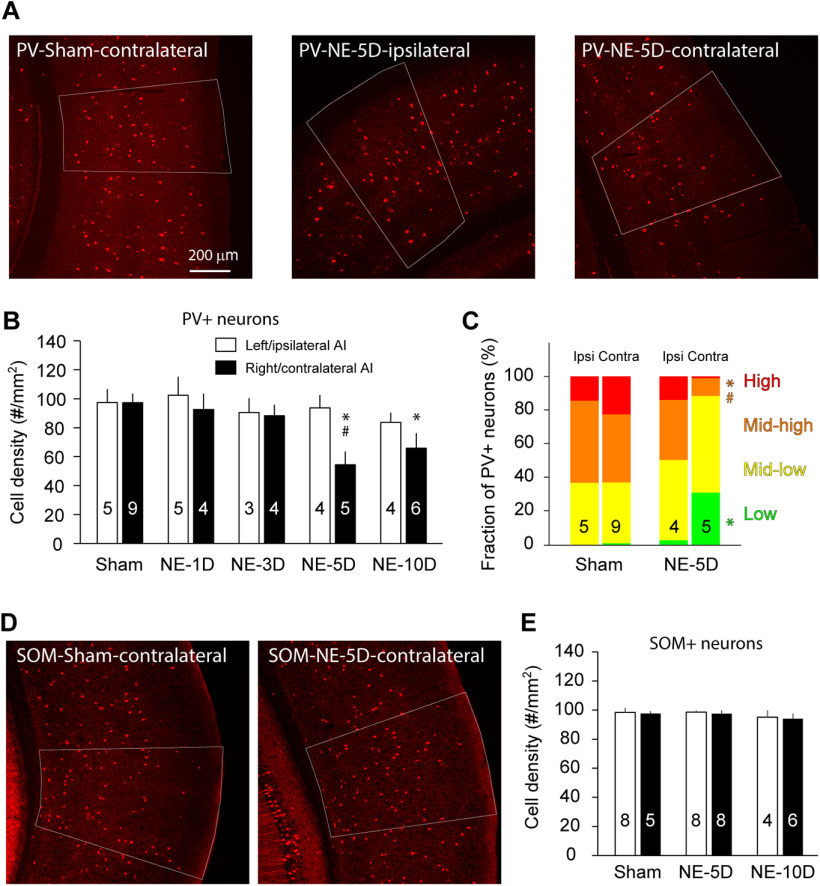
Figure 1.
Noise exposure reduces the density of PV but not SOM neurons in AI. A, Example images showing PV neurons in AI of sham (control)- and noise-exposed (NE) mice. B, Density of PV neurons in AI of control mice, and mice 1 d, 3 d, 5 d, and 10 d after noise exposure to the left ear. C, Fraction of PV+ neurons having high (>4500 a.u.), midhigh (3000–4500 a.u.), midlow (1500–3000 a.u.), and low PV (≤1500 a.u.) immunofluorescence signal levels. D, Example images showing SOM neurons in AI of sham- and noise-exposed mice. E, Density of SOM neurons in AI of control mice and mice 5 and 10 d after noise exposure to the left ear. Numbers of mice are indicated in the graphs as sample sizes. Data are presented as mean + SEM; *p < 0.05 compared with control, and #p < 0.05 compared with contralateral side.

