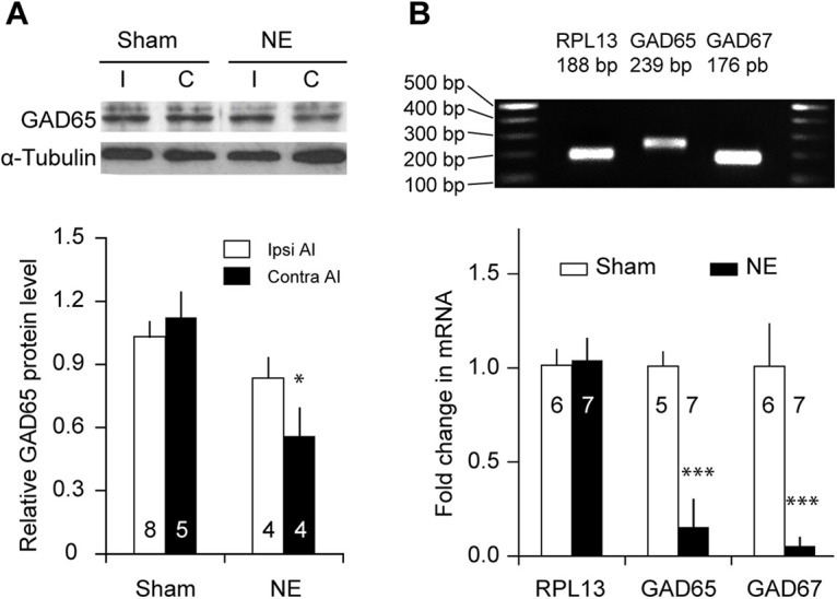
Figure 3.

Noise exposure reduces GAD expression in PV neurons. A, GAD65 expression was quantified with Western blot. The GAD65 protein level decreased significantly in contralateral AI after noise exposure (NE). α-Tubulin was used as an internal control. B, RPL13, GAD65, and GAD67 mRNA levels were measured with qRT-PCR for samples extracted from PV neurons (see above, Materials and Methods). The mean RPL13 mRNA level was unchanged by noise exposure. GAD65 and GAD67 mRNA levels were normalized to the RPL13 mRNA level for each sample. Both GAD65 and GAD67 mRNA levels were significantly reduced in noise-exposed group; *p < 0.05 compared with sham-exposed, ***p < 0.005 compared with control. Sample sizes on the graphs are numbers of mice.
