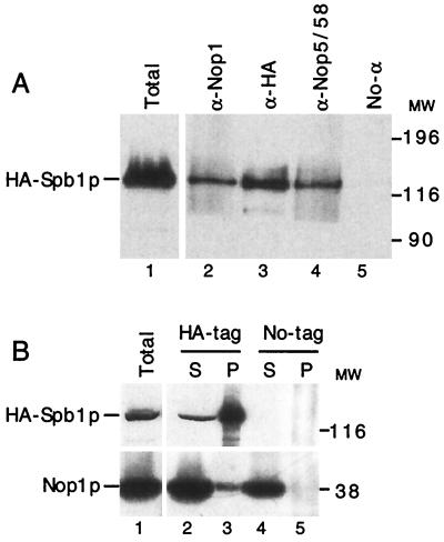
FIG. 10.
Spb1p is associated with Nop1p and Nop5/58p as shown by coimmunoprecipitation. (A) Cellular extracts prepared from a strain expressing HA-Spb1p (YDK14-1A/pDK376) were incubated with various antibodies, and the complexes were precipitated with protein A-Sepharose beads. After several extensive washes, proteins were eluted from the beads, separated by SDS–10% PAGE, and analyzed by Western blotting with anti-HA antibodies that detect HA-Spb1p. Lane 1, total cell extract corresponding to one-tenth the amount of protein used for the immunoprecipitation; lanes 2 to 5, pellets from the immunoprecipitation. Lane 2, anti-Nop1p; lane 3, anti-HA; lane 4, anti-Nop5/58; lane 5, no antibody added. MW, molecular weight markers in thousands. (B) Cellular extracts prepared from strains expressing either wild-type Spb1p or HA-tagged Spb1p were immunoprecipitated with the anti-HA antibodies. Supernatants (S) and pellets (P) were then analyzed by Western blotting with the anti-HA antibodies (upper panel) or with the anti-Nop1p antibodies (lower panel). Lane 1, total cell extract corresponding to one-tenth the amount of protein used for the immunoprecipitation; lanes 2 and 3, strain YDK14-1A/pDK423 that expresses HA-Spb1p; lanes 4 and 5, strain YDK14-1A plus pDK403 that expresses wild-type Spb1p. Lanes 2 and 4, one-tenth the supernatants; lanes 3 and 5, pellets. MW, molecular weight markers in thousands.

