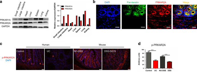
Fig. 1. Decreased phosphorylation of PRKAR2A in UC patients and mice with DSS-induced colitis.
a The expression of PRKAR1A and PRKAR2A in different tissues of WT mice. b Colon tissues of WT mice were stained with DAPI (blue), antibody to Pan-keratin (green), and antibody to PRKAR2A (red). Representative immunofluorescent images and merged images are shown. Bar = 20 μm. c Representative images of immunofluorescent staining for p-PRKAR2A(ser99) in colon tissues collected from human and mice. Nuclei were counterstained with DAPI. d Semi-quantification of the level of p-PRKAR2A in human (UC patients (n = 5) and uninflamed controls (n = 3)) and mice (treated (n = 5) and untreated (n = 5) with DSS). Data shown in a–d are representative of two independent experiments. Data are presented as mean ± SEM. Student’s t test was used to do the analysis. ***P < 0.001, bar = 50 μm.

