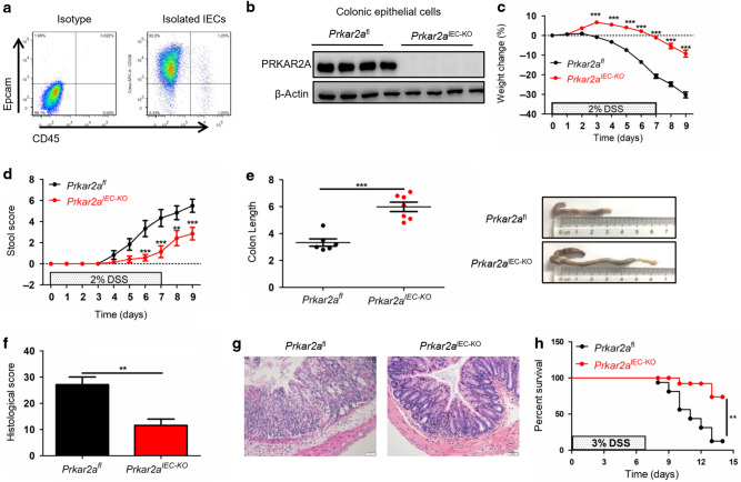
Fig. 3. Ablation of Prkar2a in IECs protects against DSS-induced colitis.
a Flow cytometric analysis of Epcam+CD45− intestinal epithelial cell frequencies in colonic epithelial cells isolated from Prkar2aIECKO mice. Cells were gated on Epcam and CD45. b Colonic epithelial cells from Prkar2aIEC-KO mice and Prkar2afl mice were collected and subjected for western blot with PRKAR2A antibody. Experiments in a, b were repeated at least three times. c–g Prkar2afl (n = 6) and Prkar2aIEC-KO (n = 7) mice were treated with 2% DSS for 1 week followed by 2 days normal drinking water. c Weight loss was monitored daily and is displayed as the percentage of the initial body weight. d Stool score was measured every day during colitis development. e At day 9, colons were removed and colon lengths were determined. f Histological analysis of distal colon tissues at day 9 of experimental colitis. g Representative images of distal colon at day 9. Scale bar = 50 μm. h The survival curve of Prkar2afl (n = 16) and Prkar2aIEC-KO (n = 12) mice. Mice were treated with 3% DSS for 1 week and the mortality was monitored over 14 days. Log-rank (Mantel–Cox) test was used to do the analysis. Data shown in c–h are representative of three independent experiments. All graphs show mean ± SEM. Student’s t test (e, f) or two-way ANOVA (c, d) was used to compare experimental groups. ***P < 0.001; **P < 0.01.

